Age related macular degeneration (AMD)
Early AMD - with low, medium or high risk of progression. Usually not a/w visual disturbances Late AMD:
- Dry - well defined areas of atrophy of retinal pigment epithelium, drusen pigment changes 90% of cases. Has a chance to progress into wet AMD
- Wet - characterised by choroidal neovascularisation, 10% of total AMD cases
Causes/Factors
- Most commonly age-related - in people aged 50 and over
- Can also be due to Diabetes Mellitus, Nutritional disorders, and genetics
Symptoms
- Distortion of vision, where straight lines appear crooked or wavy.
- Painless loss, or blurring, of central or near-central vision.
- A black or grey patch affecting the central field of vision (scotoma).
- Difficulty reading, driving, or seeing fine detail (such as facial features).
- Flickering or flashing lights (photopsia).
- Difficulty adjusting from bright to dim lighting.
- Visual hallucinations (associated with severe visual loss).
- Visual distortions - straight lines may appear wavy or bent
Signs
-
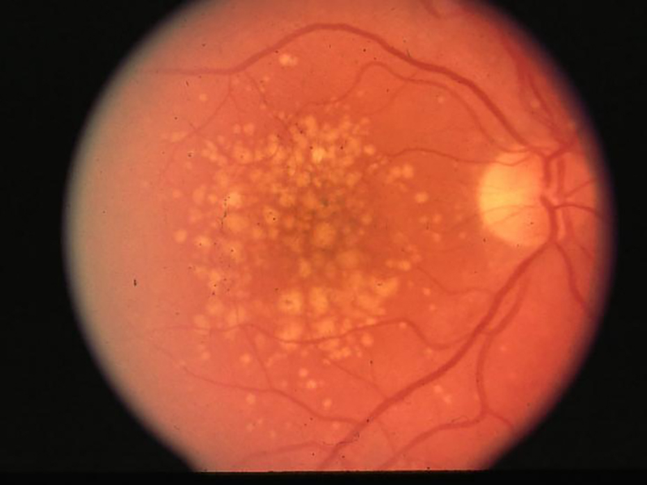
Drusen - collections of lipid material that accumulate beneath the retinal pigment epithelium (RPE) and within Brunch’s membrane
-
Retinal pigment epithelial changes
-
Geographic atrophy
-
Choroidal neovascularisation - abnormal blood vessel growth beneath the retina in wet macular degeneration
Investigations
- Retinal exam - dilated fundus exam
- Optical coherence tomography (OCT) high resolution imaging to assess retinal thickness and integrity
- Fluorescein angiography - to detect choroidal neovasculatisation in wet macular degeneration
Management
Dry AMD
- Not much is usually done
- Lifestyle factors stop smoking & controlling blood pressure
- Some studies have shown vitamin supplementation to be effective
Wet AMD
- To try and prevent neovascularization, anti-VEGF (vascular endothelial growth factor) can be injected in the vitreous about once a month
Complications/red Flags
- Visual impairment and blindness.
- Visual hallucinations.
- Depression.
- Falls and fractures.
- Reduced quality of life