Most common forms are diabetic retinopathy, diabetic macular oedema and Cataracts
Diabetic retinopathy
-
Most common diabetes related complication.
-
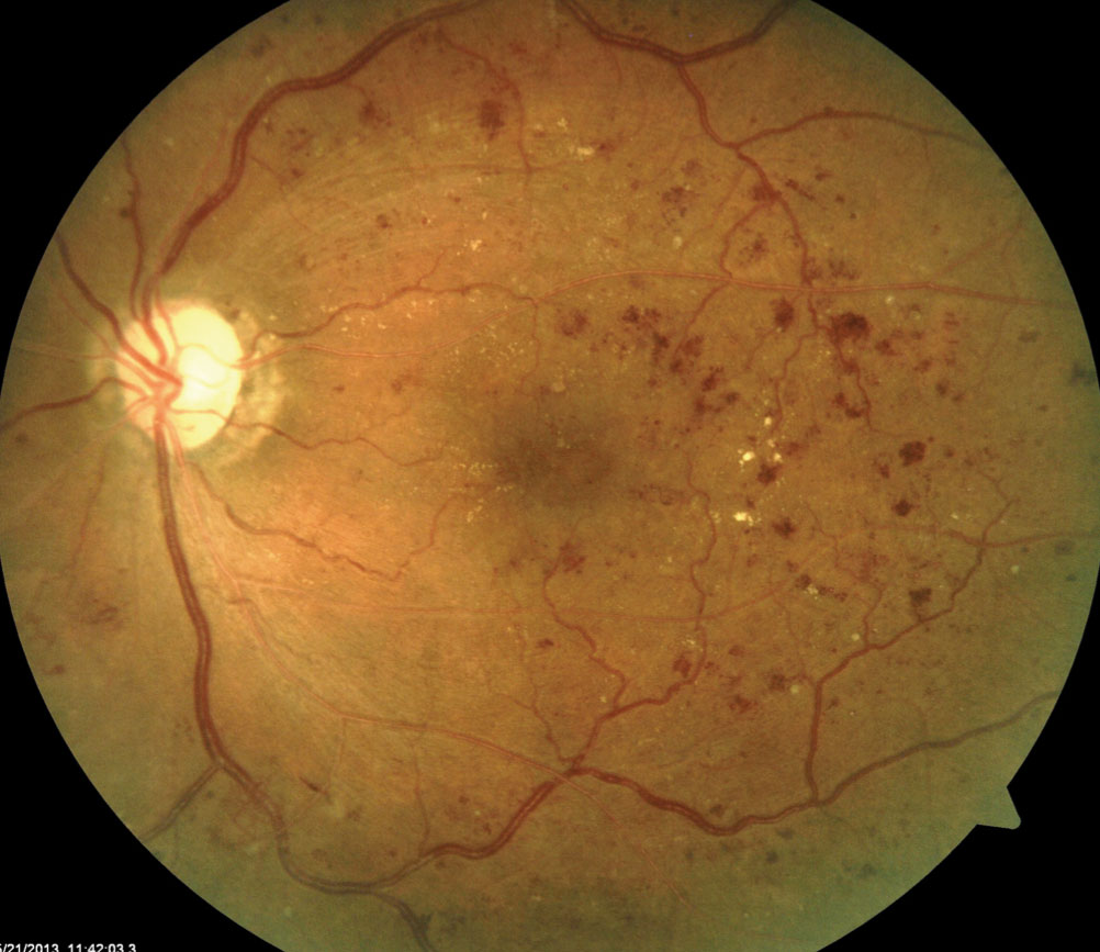
Intraretinal microvascular abnormalities (IRMA) - dilated and tortuous capillaries that can act as a shunt
-
Neovascularization - release of growth factors stimulating new vessel development - proliferative diabetic retinopathy
-
Damage to the wall of small vessels cause microaneurysms.
-
Damaged blood vessels leak fluid into the retina. This fluid leaves behind lipids and proteins forming hard exudates
-
Damage to nerve fibres in to the retina causes cotton wool spots.
-
These can all be seen on dilated eye exam.
-
Fluid from leaking vessels in cleared poorly in the macular area due to anatomical differences. Above a certain point it cannot be cleared macular oedema occurs
-
This distorts and thickens the macula at the retina.
-
Not visible on exam
If proliferative (neovascularization occurs), measures taken to treat:
- Pan-retinal photocoagulation (PRP) – extensive laser treatment across the retina to suppress new vessels
- Anti-VEGF medications by intravitreal injection
- Surgery (e.g., vitrectomy) may be required in severe disease
Cataracts
- Develops earlier in people with diabetes tan the general population
- Fluctuations in blood glucose concentration can cause refractive variability as a result of osmotic changes within the lens
- Resolves with better control
Presentation
- Chronic visual acuity loss - finding it hard to read
- Colours having a grey/brown/yellow tinge
- Star-bursts around lights
Causes/Factors
- Diabetes Mellitus: Prolonged elevated blood sugar levels contribute to the development and progression of diabetic eye disease.
- Duration of Diabetes: The longer an individual has diabetes, the higher the risk of developing eye complications.
- Essential hypertension: High blood pressure can exacerbate the effects of diabetes on the eyes.
Symptoms
- Blurred Vision: Vision may become blurry, especially in the early stages.
- Floaters: Dark spots or floaters may appear in the field of vision.
- Eye Pain or Pressure: In advanced stages, individuals may experience pain or pressure in the eyes.
Signs
- Retinal Changes: Fundoscopic examination may reveal abnormalities in the retina, such as microaneurysms or haemorrhages.
- Macular Oedema: Swelling of the macula, the central part of the retina, can be a sign of diabetic macular oedema.
- Cataracts: Clouding of the eye’s lens.
- Increased Intraocular Pressure: A risk factor for glaucoma.
Diagnostic Tests
- Dilated Eye Exam: An eye care professional examines the retina and other structures after dilating the pupils.
- Fluorescein Angiography: A dye is injected into the bloodstream to highlight blood vessels in the retina.
- Optical Coherence Tomography (OCT): Imaging technique to assess the thickness of the retina and detect swelling.
Management
- Blood Sugar Control: Tight control of blood glucose levels is crucial to prevent and manage diabetic eye disease.
- Blood Pressure Management: Maintaining optimal blood pressure helps protect the eyes.
- Anti-VEGF Injections: Injections may be used to treat diabetic macular edema.
- Surgery: Advanced cases may require surgery, such as vitrectomy for retinal issues.
Complications/Red Flags
- Vision Loss: Progressive vision loss can occur if diabetic eye disease is not adequately managed.
- Retinal detachment: In severe cases, the retina may detach.
- Glaucoma: Increased risk of developing glaucoma, leading to optic nerve damage.